Membranes are extremely thin - only 4 to 5 nanometers thick - and very dynamic. Even the tiniest changes can have a big impact. For example, a small change in the chemical structure or an exchange of salts (electrolytes) can greatly affect the properties of the membrane.
It is therefore important to know exactly what a membrane is made of and to be able to measure changes in it. This is where nanoanalytics comes in. This requires a combination of different analytical methods, such as thin-layer chromatography, differential calorimetry or fluorescence spectroscopy. Our special method is small-angle X-ray and neutron scattering. Using this technique, we have developed our own models over the years to analyze membranes even better.
Combining X-Ray and Neutron Scattering Provides More Detailed Insight
Small-angle X-ray and neutron scattering (SAXS, SANS) can visualize structures in the size range from about 1 to 100 nm - ideal for studying membranes. By combining the two methods, we can determine details such as layer thickness or the space occupied by a lipid in the inner and outer leaflets of the membrane separately. This is especially exciting for asymmetric membranes. It also allows us to determine the molecular shape of lipids and to observe how membrane-active peptides are inserted into the membrane.
Publications:
- Semeraro et al., in Methods in Enzymol, T. Baumgart, M. Deserno (edts), Academic Press, 700: 349 - 383 (2024) DOI: 10.1016/bs.mie.2024.02.017
- Semeraro et al., Soft Matter 17: 222 - 232 (2021) DOI: 10.1039/C9SM02352F
- Kaltenegger, et al., Biochim. Biophys. Acta 1863: 183709 (2021). DOI: 10.1016/j.bbamem.2021.183709

Design: G. Pabst
Examples
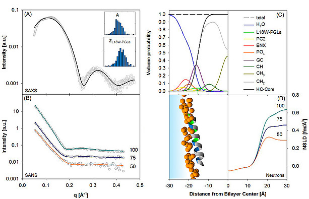
Design: G. Pabst, data from Pachler et al., Biophys J (2019) , License: (CC-BY 4.0 DEED)
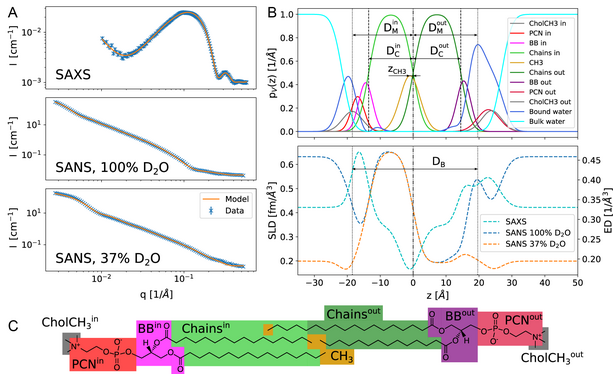
From: Frewein et al, J Membrane Biol (2022), License: (CC-BY 4.0 DEED).
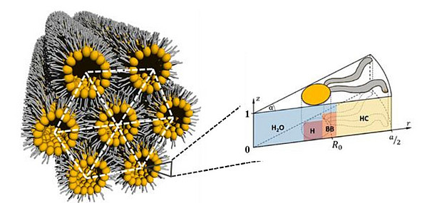
Design: G. Pabst, M. Frewein