Research
Neuroanatomy of the spinal cord
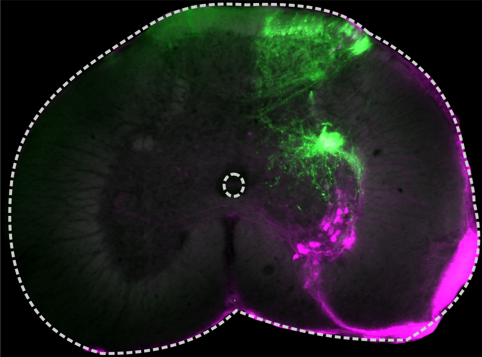
Semi-intact, in-vitro tissue can be used to perform staining of whole spinal nerves. Such staining allows to trace neurons that project through the respective nerves, including their dendritic and terminal fields. The data can be used to describe the connectivity and distribution e.g., of sensory input- and motor output layers in the spinal cord. The displayed image shows a staining of the sensory input- and the output layers of the spinal cord of a rattlesnake. Through tracer application on ventral and dorsal roots, motoneurons (magenta) and terminals of sensory neurons (green) were visualized.
Pectoral motor pool organization in teleosts
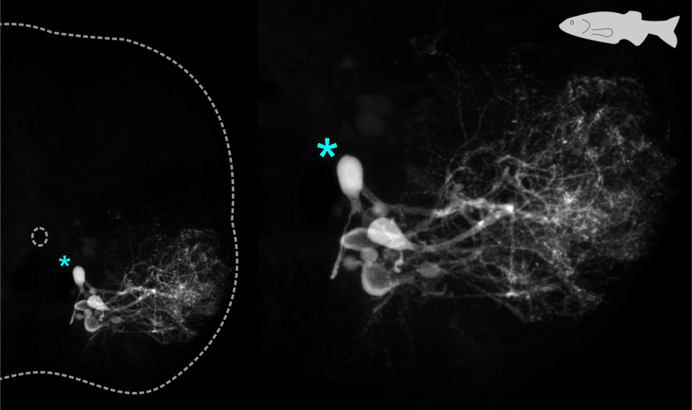
Injections of neural tracers into specific muscles can be used for targeted backfills of motoneurons that innervate them. In combination with investigations of motoneuron electrophysiology, this gives important insights into the anatomical and physiological organization of motor pools within the spinal cord. One of my research interests lies in the comparative analysis of such data recorded from different teleosts that perform specialized pectoral motor behaviors. The insight gained from such comparative studies can provide important learnings on the organizational principles of motor pools that allow for the targeted activtion of specific muscles by the central nervous system to execute different motor behaviors.
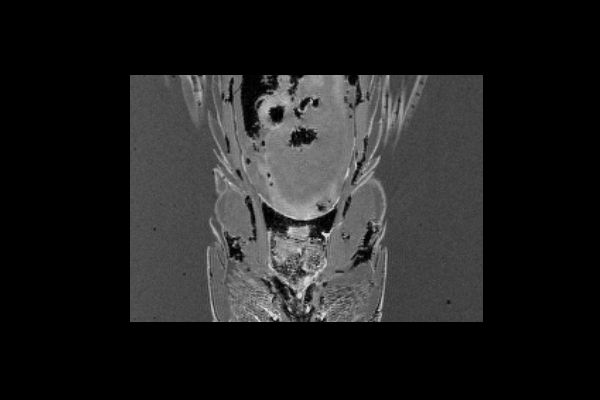
µCT and MRI imaging of teleost musculoskeletal systems
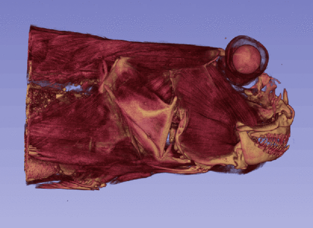
The University of Graz offers fascilities and experienced staff to perform micro-computed tomography (µCT) and magnetic resonance imaging (MRI) of many types of samples. Combining µCT and MRI enables detailed reconstructions of musculoskeletal systems of fish. Unstained specimen can be scanned by means of µCT to provide images of the skeleton. Incubation of specimen in iodine-solutions such as Lugol’s, allows for detailed µCT scans of soft tissue. This is due to the incorporation of iodine into soft tissues which greatly enhances tissue contrast for µCT scans. MR imaging provides additional information, for example, on muscular structure by methods like diffusion tensor imaging (DTI). Posthoc aligment allows for an integrated analysis of the different scans and musculoskeletal modelling. Open source software packages like 3D slicer can be used to extract the 3D volume of individual structures of interest from scans by means of different approaches, including manual and threshold-based segmentation.
Behavioral tracking
Markerless motion tracking
To allow for a thorough interpretation of experimental results from investigations of neural networks that underlie locomotor behavior of animals, it is important to integrate data on neurophysiological and anatomical characteristics of motor networks with data on the actual execution of the motor behaviors they control. In recent years, great progress has been made in the development of markerless motion tracking: a ready to use software package for such tracking analysis is DeepLabCut. It provides a ready to use interface that allows researchers to train neural networks to perform reliable and fast tracking of predefined body parts from video recordings. Tracking data is afterwards available for further analysis.
To analyze the kinematics of motor behaviors, I record freely moving animals and use DeepLabCut for automated tracking of bodyparts involved in the execution of locomotor patterns. The integrated analysis of motor behavior kinematics, associated musculoskeletal systems and anatomical and physiological characteristics of neural networks underlying the execution of motor behaviors, provides great chances to get deeper insights into the adaptability of motor programs and which anatomical and physiological organization principles are associated with it.
To the right, you can see data from a preliminary analysis of locomotor behaviors of an african mudskipper while swimming under water (left panel) and while performing crutching locomotion on land (right panel). For swimming, the amplitude of motion of tracked bodyparts increases the further caudal they lie. Also note the phase shift of undulatory motion along the rostrocaudal body axis, indicating sequencial muscle activation. The right panel shows the execution of one "step" of the animal on land. Here, markers are set to track the pectoral and pelvic fin, as well as a point on the upper back of the animal.
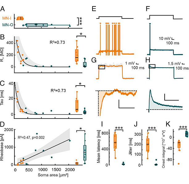
Electrophysiology
Using patch clamp recordings of motoneurons allows for detailed investigations of their electrophysiological characteristics. Manipulation e.g., by means of pharmacologically blocking specific types of ion channels, allows for insights into the underlying molecular basis of motoneuron electrophysiological properties.
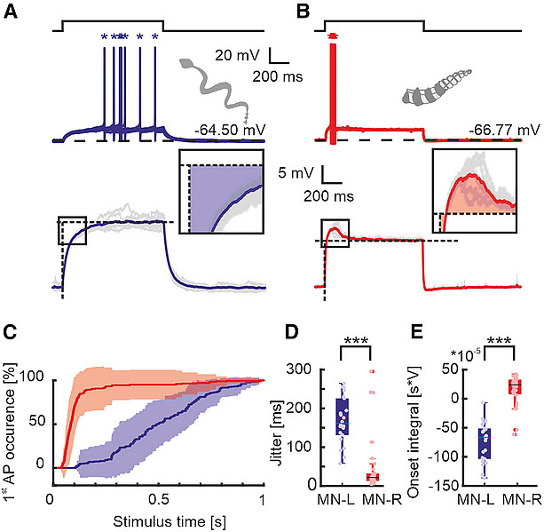
Bothe, M. S., et al. (2024). Timing and precision of rattlesnake spinal motoneurons are determined by the KV72/3 potassium channel. Current Biology, 34(2), 286-297.e5., licence: CC BY 4.0
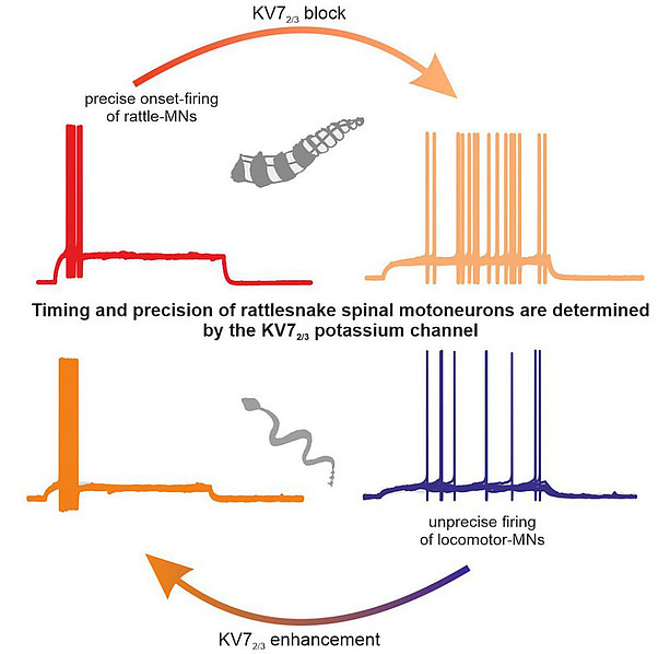
Bothe, M. S., et al. (2024). Timing and precision of rattlesnake spinal motoneurons are determined by the KV72/3 potassium channel. Current Biology, 34(2), 286-297.e5., licence: CC BY 4.0
*Gutjahr, R., *Bothe, M. S., et al. (2024). Diversification of pectoral control through motor pool extension. Proceedings of the National Academy of Sciences, 121(49). *Equal contribution. Licence: CC BY-NC-ND 4.0