Research interests
BioImaging Lab
At the Bioimaging Lab, we combine experimental life sciences, modern microscopy and computer-aided image analysis to investigate biological systems across a range of scales, from bacteria and yeast through to Drosophila, and on to mammalian cells and tissues. As part of our own research and collaborative projects both within and outside the University of Graz, our activities encompass five interlinked areas, ranging from fundamental cell biology and the development of advanced imaging strategies, through quantitative bioimaging approaches and bespoke AI-based analyses, to open and reproducible science.
01 Lipid droplets at the interface with the cell nucleus
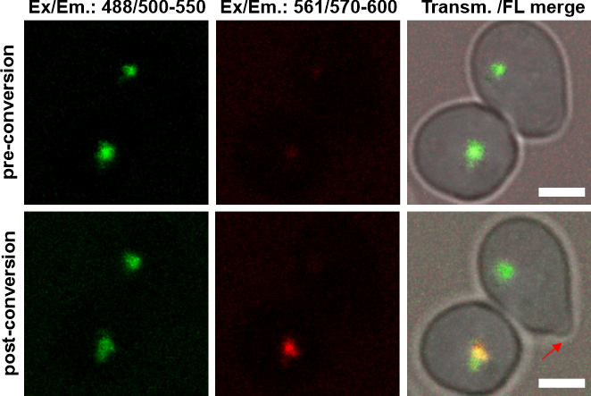
Lipid droplets are highly dynamic organelles and central hubs of cellular lipid and energy metabolism. They store neutral lipids, provide lipids for energy production and membrane biogenesis, and contribute to protecting cells from lipotoxicity and metabolic stress. Their formation, composition, and interactions with other organelles are dynamically adapted to the physiological state of the cell. A particular focus of our research is the seipin complex, a key regulator of lipid droplet formation, and the still poorly understood relationship between lipid droplets, the nuclear envelope, and nuclear lipid homeostasis. The physiological importance of seipin is particularly evident in humans: mutations in the BSCL2 gene, which encodes seipin, can cause severe congenital generalized lipodystrophy, in which neutral lipid storage in adipose tissue is profoundly impaired and serious metabolic complications can develop. Understanding the fundamental cellular functions of seipin and the lipid droplet processes it controls is therefore also important for elucidating disease-relevant mechanisms. In addition to molecular and cell biological approaches, 4D live-cell microscopy is an important experimental strategy for directly investigating dynamic and potentially heterogeneous processes in individual living cells. Using photoconvertible markers, defined LD subpopulations can be followed across different growth phases and throughout the cell cycle, allowing their individual fates to be tracked. This approach aims to determine how distinct LD populations arise, change over time, and contribute to cellular and nuclear lipid homeostasis, as well as how disturbances in these processes may contribute to the development of disorders such as lipodystrophy.
02 New approaches to the quantitative imaging of lipid droplets
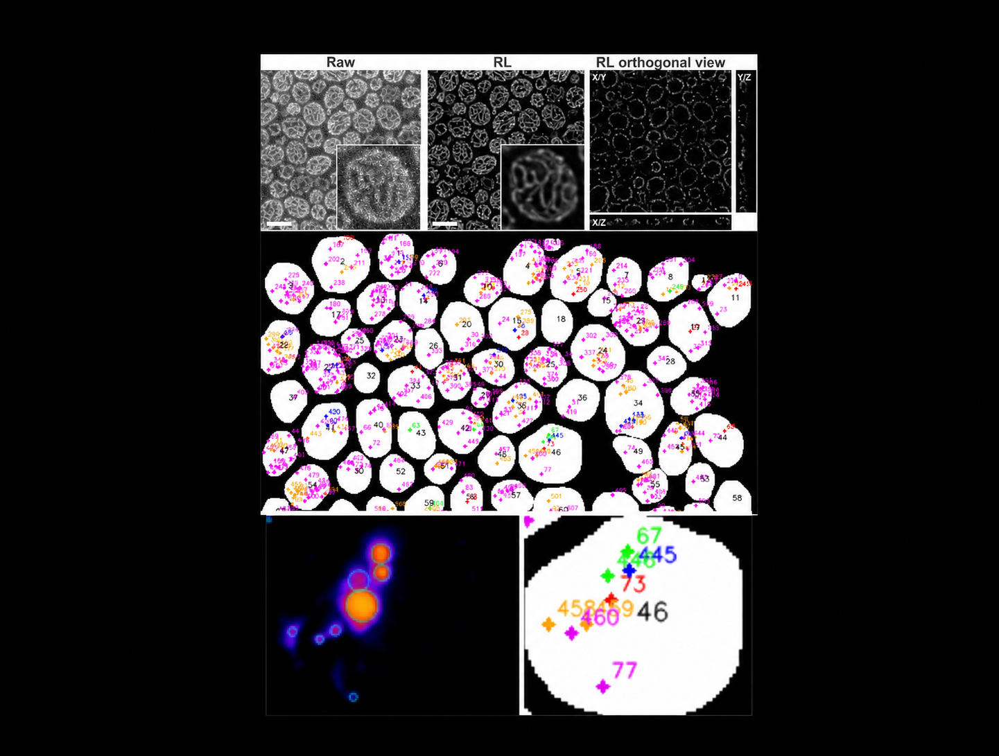
Answering such biological questions requires not only dynamic imaging but also statistically robust morphometric analyses of large cell populations. To this end, an experimental platform for the quantitative analysis of lipid droplets has recently been established, which links biological parameters to the physical fundamentals of microscopic imaging. At the same time, this work demonstrated the extent to which quantitative results can be influenced by image acquisition, processing and the method of analysis. Building on this, strategies are currently being developed to quantify methodological limitations and to validate quantitative bioimaging methods more objectively.
03 Tailor-made deep learning for bioimage analysis
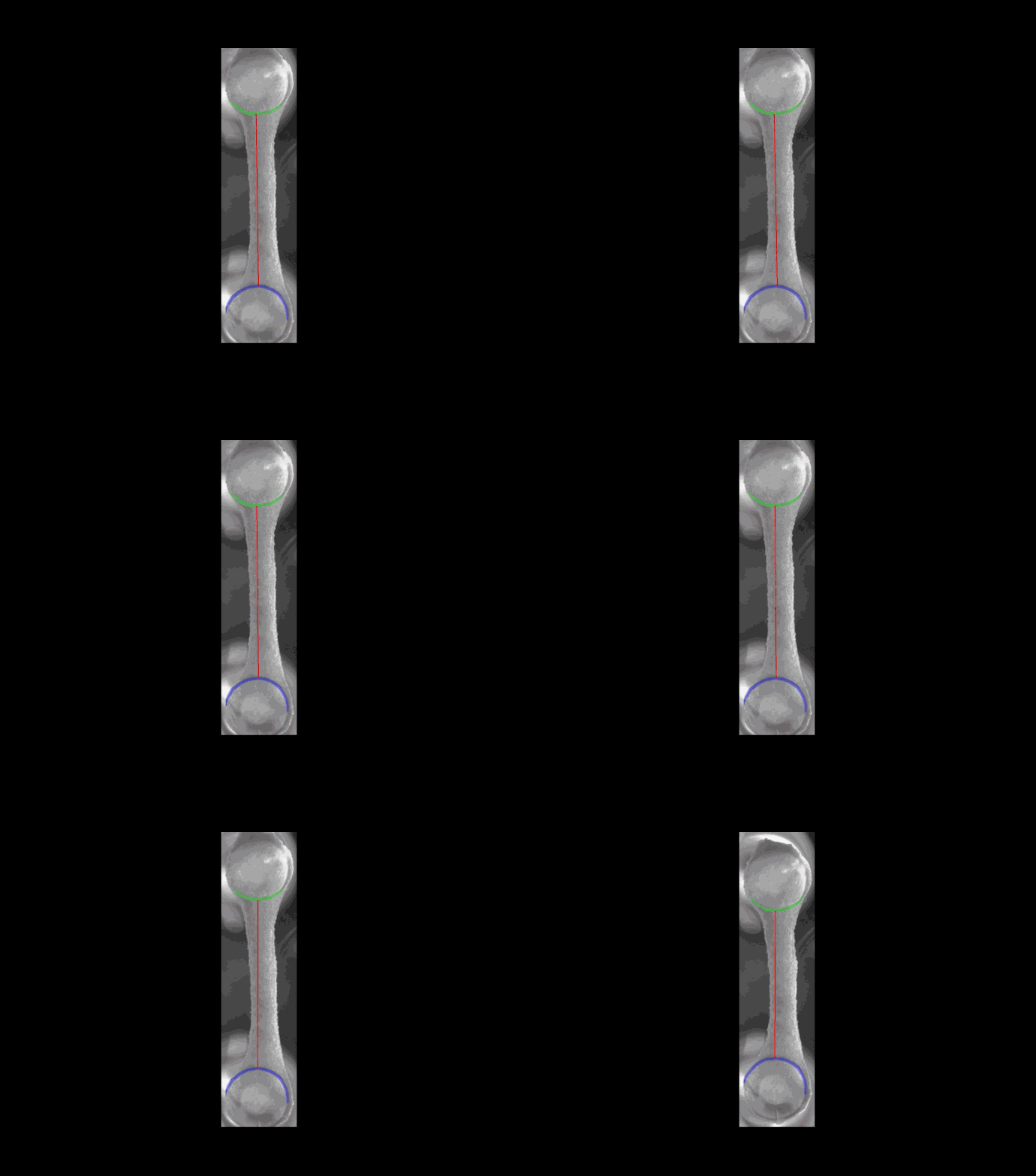
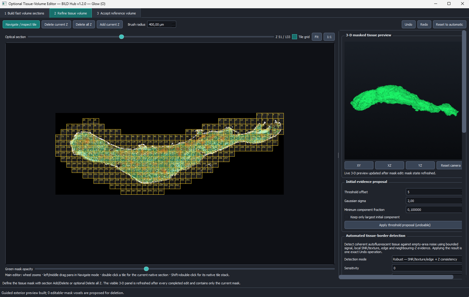
We have developed our own modular deep learning platform for biological image data, which can be flexibly adapted to different research questions and sample types. The “BioImaging Lab Deep Learning Hub (BILD-HUB)” combines application-specific training, segmentation, validation and quantitative analysis, and is already being used for various applications, including the analysis of contraction frequencies in engineered heart tissue (EHT) and the continuation of our work on the 3D quantification of neuronal structures in mouse adipose tissue (Rauchenwald et al. 2025. JCS 138(3):JCS263438). Furthermore, the platform expands our research into lipid droplets and seipin and opens up new possibilities for analysing complex LD phenotypes and subpopulations.
A key objective of our analysis platforms, however, is not only the quantitative evaluation of biological image data, but also the systematic comparison of different analytical approaches in order to identify and quantitatively assess quantification errors and methodological biases.
04 3D imaging of complex tissues
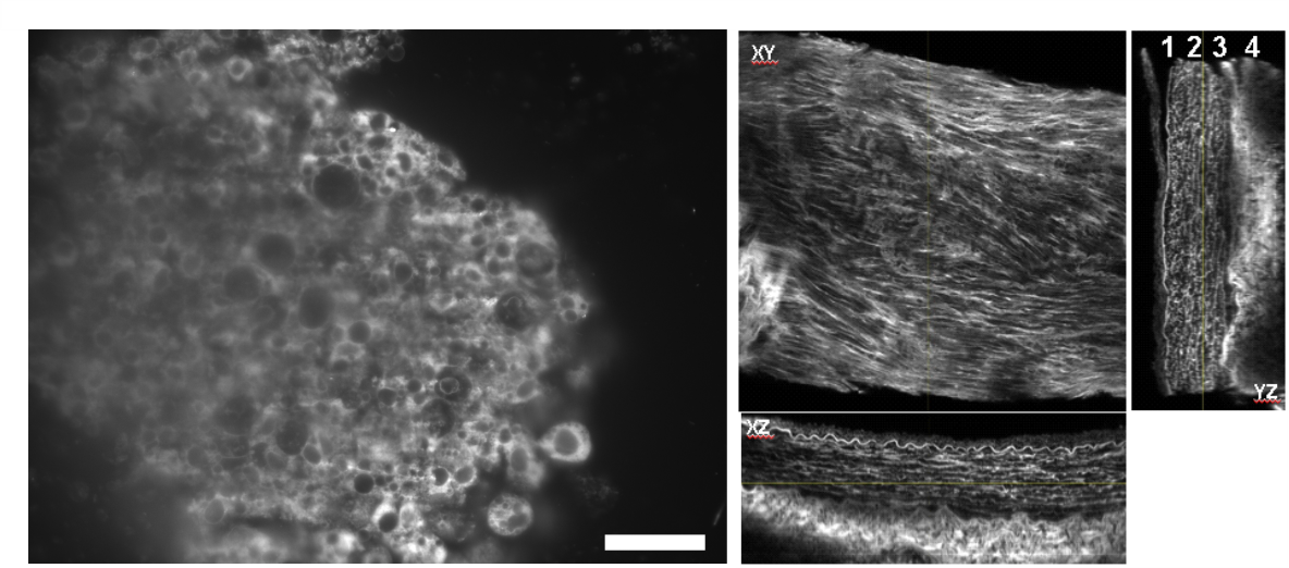
Reliable quantitative analysis begins with high-quality raw data. A further focus is therefore on the development and optimisation of sample preparation and imaging techniques for complex three-dimensional biological systems. These include, in particular, optical tissue clearing, high-resolution confocal and light-sheet microscopy, and their adaptation to different samples. Such strategies have, for example, been established for the high-resolution visualisation of collagen architecture in optically cleared rabbit aortas. These approaches are currently being applied to other 3D models such as spheroids. In the future, they are also intended to be used to investigate seipin, lipid droplets and their spatial organisation in more complex tissues and model systems. In this context, sample preparation, image acquisition and computer-aided analysis are understood as an integrated experimental workflow.
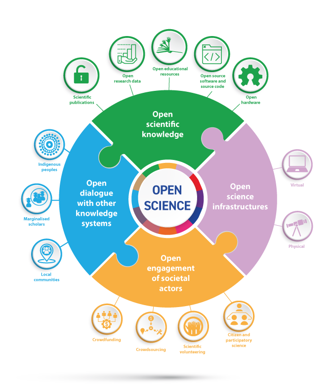
05 Beyond ‘Black Boxes’: Open Science as a guiding principle
For us, Open Science is not an option to research, but an integral part of its design. Our aim is to make methods, code, data and analytical decisions accessible, transparent and reproducible, as we have already done in our current work on quantitative bioimaging.
By openly documenting both the possibilities and the limitations of our approaches, we aim to help make quantitative imaging in the life sciences more easily reproducible, verifiable and further developable. Through these activities, we are also contributing to the University of Graz’s broader open science initiatives and supporting a research culture in which experimental data and methods can be traced, tested and reused, rather than being treated as ‘black boxes’.
H. Wolinski. 2026. Open Science in Quantitative Bioimaging – An Oxymoron? In: Open Science – Opportunity or Challenge. Conference proceedings. doi.org/10.25364/978-3-903374-47-8